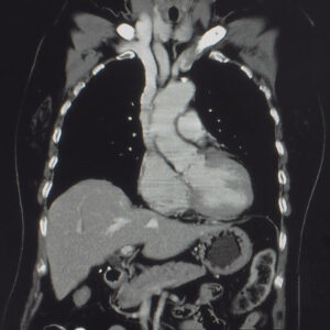
Computed tomography (CT) is a non-invasive, detailed series of images that combines a series of X-rays taken at different angles to create cross-section images. These images can be viewed in two and three dimensions. Ionising radiation is used to identify abnormalities by producing diagnostic, high-resolution images of the body’s internal structures. CTs are also used as guidance for certain procedures such as biopsies, placement of drains and infiltrations (interventional radiology).
During CTs patients are required to lie down on a bed in a doughnut-shaped machine with a short tunnel in the centre. Patients need to remain completely still for the duration of the scan because movement will compromise the quality of the scan. Although CTs are painless, patients with injuries or painful clinical conditions may experience some discomfort. Communication with the radiographer is possible via an intercom.
Some CT scans require oral and/or intravenous contrast. The intravenous contrast is iodine based. Contrast media is used to optimise the visualisation of organs, structures, and vessels in the body. The radiologist or radiographer will insert an intravenous catheter (IV line) into a vein in the patient’s hand or arm. Contrast injections may cause a flush of heat and/or a metallic taste. These sensations usually disappear within a minute or two. At some stages of the scan, patients may be required to breathe as instructed.
