Milpark

Mammography

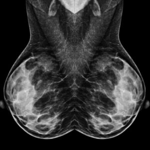
Mammography is the gold standard imaging modality to examine breast tissue and uses a very low dose of ionising radiation. Early detection and diagnosis of breast diseases in both women and men are essential for effective treatment. 

Mammograms are recommended annually for women from the age of 40 years. High-risk patients, usually those with a family history, should start screening 10 years earlier than the age of the diagnosis of a first-degree relative such as a mother or sister. Alternatively, high-resolution ultrasounds will be performed for younger patients. All patients, including those younger than 40 years, should do monthly self-examinations. Patients with symptoms such as a lump in the breast, nipple discharge, pain, and changes in the shape or texture of the nipple or breast should have breast imaging done as soon as possible. 

Digital breast mammograms with tomosynthesis are a detailed and advanced form of breast imaging available at our practice and recommended for dense breasts. Tomosynthesis is 3D imaging where multiple images of different angles of the breast tissue are captured and reconstructed. This X-ray involves compression of the breast tissue which may cause temporary discomfort for patients that have a painful clinical condition or sensitive breasts.

Mammograms are routinely complemented by high-resolution breast ultrasounds. For breast ultrasounds, a water-based, easy to clean gel will be applied to the breasts and axilla/armpit. A device called a transducer is used to obtain the images. 

Our radiologists and mammographers are specially trained to provide these services in a relaxing, supportive, and professional environment.

We offer the following imaging services:

  • Collection/abscess drainages
  • Core biopsies – ultrasound and stereotactic guidance
  • Mammography with tomosynthesis (3D imaging)
  • Tissue marker placements
  • Ultrasounds
  • Vacuum-assisted biopsies
Mammography

Please note:

  • Patients are responsible for obtaining authorisation for radiology procedures. Please contact us for assistance.
  • Women must inform their referring clinician and the mammographer on duty if there is any possibility that they may be pregnant. 
  • Patients must inform the mammographer of any symptoms, prior surgeries, or breast prostheses.
  • For comparison and accurate diagnosis, patients must bring previous mammogram images and reports to their appointment.
  • Patients will be asked to change into a hospital gown and to remove jewellery and other metallic items from their body.
  • Should a core biopsy be required, patients must inform the radiologist or mammographer if they are using any blood thinning medication or have any allergies. Specific blood tests may be required. Radiologists take special care to explain the procedure and the necessary post-care with each patient, ensuring that they are comfortable and understand what it entails.

Frequently asked questions

For breast screening benefits, please contact your medical aid.

Do not panic. Contact our practice and schedule an appointment. We will evaluate and perform the necessary breast imaging depending on your age and symptoms. 

Yes, mammograms with compression are safe for patients with protheses.

Mammograms and ultrasounds imaging predict possible breast cancers. To confirm the diagnosis, a tissue biopsy is required. 

A biopsy is a sample of tissue taken from the breast for analysis and diagnosis. The procedure is done by the radiologist, and the tissue is evaluated by a pathologist.

On average, three to four working days. The results will be sent to your referring clinician.